The world’s top 2% of most impact scientists across all fields have been updated in a new list issued in October 2023 by Stanford University (1). Dr. Teodor Parella, head of the SeRMN-UAB, appears in this Stanford list that includes 3230 spanish researchers from all disciplines, around 300 spanish chemists and 81 researchers from the Universitat Autònoma de Barcelona. Dr. Parella is found at the top world-wide 1% scientists list, ranking #80088 in the world, #980 in Spain and #21 at the UAB.
Dr. Parella has also been ranked #6 at UAB, #268 in Spain and #7687 in the world in the field of Chemistry, according toResearch.com, a leading academic platform for researchers, that has just released the 2023 Edition of the Ranking of Best Scientists. The ranking is based on D-index (Discipline H-index) metric, which only includes papers and citation values for an examined discipline. The ranking includes only leading scientists with D-index of at least 40 for academic publications made in the area of Chemistry.
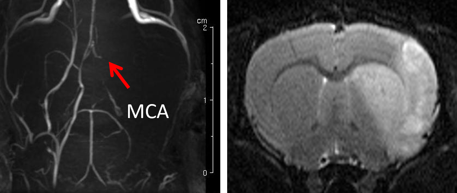
Last 26th July 2023 I successfully defended my PhD Thesis entitled: “PhD Thesis: Implementation of high-resolution MRSI methods in a pre-clinical scanner, and optimization for brain longitudinal studies of therapy response in mice glioma model.”
Abstract
Magnetic resonance spectroscopy and magnetic resonance spectroscopic imaging (MRS/MRSI) are non-invasive diagnostic techniques that use a strong magnetic field and radio waves to examine the chemical composition of living tissue. Working on the same principles as Magnetic Resonance Imaging (MRI), instead of producing images, MRS generates a spectrum of signals that can be used to identify the type and amount of molecules present in a tissue. The utility of MRS and MRSI has already been established in many studies, providing useful information about the chemical makeup of different regions of the brain, and allowing diagnosis of conditions such as Alzheimer’s disease, multiple sclerosis, and brain Glioblastoma (GB) tumors.
Preclinical glioblastoma studies looking forward to improving therapeutic outcomes are necessary since clinical GB has no current cure. These studies can greatly benefit from improved spatial resolution and homogeneity of the acquired MRSI grids. Hence, we can work towards improved acquisition schemes enhancing the quality of acquired data using MRS and MRSI. There exists a methodological consensus among spectroscopy experts where the Localized Adiabatic Spin Echo Refocused (semiLASER) data acquisition strategy has been ranked as the most likely localization technique to improve (pre) clinical MRS. SemiLASER uses adiabatic pulses to selectively excite and refocus the signal from a localized volume of interest in the brain. This results in a higher signal-to-noise ratio (SNR) and better spatial resolution compared to conventional data acquisition sequences.
Partial volume effects can occur in MRSI when the voxel (a 3D volume of interest) being measured contains a mixture of different neighbouring tissue types or compartments, such as grey and white matter or cerebrospinal fluid. This can lead to inaccurate quantification of metabolites, as the signal from one tissue can mix with the signal from another and affect overall pattern recorded. SemiLASER is designed to minimize partial volume effects by using adiabatic pulses to selectively excite and refocus the signal from a small region of interest within the voxel. This allows for more accurate quantification of metabolites within the region of interest, while reducing the contamination of the signal by other tissue types. In addition, semiLASER also employs an outer-volume suppression (OVS) technique to further reduce contamination from outside the region of interest. This involves using additional adiabatic pulses to selectively saturate the signal from outside the volume of interest, so that it does not contribute to the MRSI signal. Overall, the combination of selective excitation and OVS in semiLASER can help improve the accuracy of MRSI measurements and reduce partial volume effects.
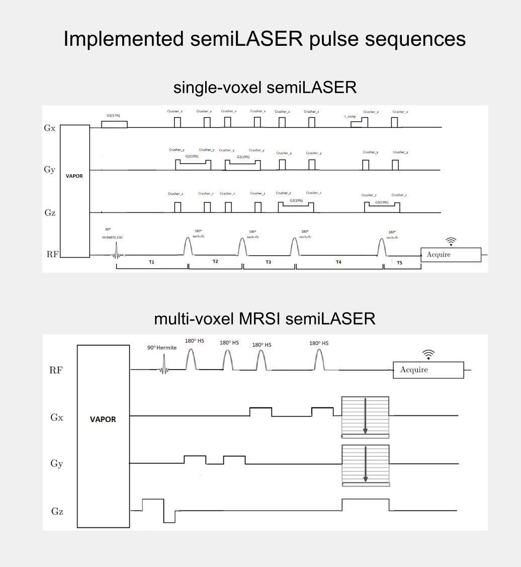
Although, the clinical utility of semiLASER has been acknowledged, the pre-clinical use and implementation of semiLASER still remains a less explored area. Our group has a long record of using MRSI in therapy response monitoring of a murine model glioblastoma (the GL261 cell line) using a commercially available MRSI acquisition sequence. In our efforts towards bridging the barriers between pre-clinical and clinical research, we have implemented the clinically verified semiLASER sequence on a pre-clinical 7T Bruker Biospec USR scanner running the ParaVision 5.1 software package, which provides a graphical user interface for sequence programming and data acquisition. The single and multi-voxel semiLASER sequences were implemented with the idea that the developments generated during this PhD project will be replicable by other interested users.
The implemented SV-semiLASER and MRSI-semiLASER sequences for preclinical acquisitions were optimised to perform high resolution MRSI of living mouse brain. For this, sequences were duly verified and tested first in phantoms and later in-vivo, in wild-type (wt) and tumor bearing (GL261) mice. To do so, the Bruker pulse sequence implementation was first studied in detail to become familiar with the Bruker programming environment and a test sequence PRESS_Slice to localize the slice dimension was developed by modifying the Bruker stock PRESS sequence for single voxel localization. After careful evaluation of test sequence results, the semiLASER single and multi-voxel sequences were also implemented using a similar strategy.
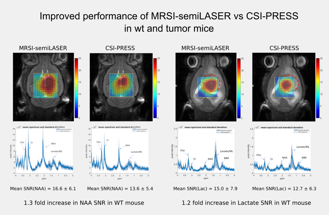
The implemented SV-semiLASER sequence provided a ca. 1.4-fold improvement in SNR in phantoms and ca. 1.3-fold improvement in SNR for in-vivo subjects, in comparison to the stock Bruker PRESS (single volume acquisition) sequence. The MRSI-semiLASER sequence resulted in a ca. 1.3-fold improvement of SNR in phantoms and in-vivo subjects compared to the stock Bruker CSI-PRESS sequence. Combined with phase encoding strategies and volume reduction methods, higher spatial resolution and SNR was achieved for the implemented MRSI-semiLASER. The quantification analysis of the results was done using MATLAB based post-processing tools specially designed to process Bruker datasets and solutions for a faster post processing pipeline were proposed. The single voxel MRSI-semiLASER sequences were further simulated using NMRSCOPE-B virtual simulator, a jMRUI plug-in which positively correlated with the experimental results. Preliminary nosological images obtained using MRSI-semiLASER datasets and the SpectraClassifier tool previously developed in our group, and trained with GL261 tumors using already available CSI-PRESS data, suggested those classifiers could be robust enough to recognize the tumor region acquired with the semi-LASER sequence. Still, classifiers may require retraining for the evaluation of response to therapy, which is an ongoing project within the group.
The thesis dissertation can be downloaded in PDF format using the link below or from the official TXD and Teseo repositories (currently in progress):
I would like to thank the financial support by the European Comission Marie Curie Initial Training Networks (ITN, call H2020-MSCA-ITN-2018, grant 813120 to project INSPiRE-MED); by the Ministry of Science and Insdustry (MCIN/AEI/10.13039/501100011033) (APC); and by Centro de Investigación Biomédica en Red—Bioingeniería, Biomateriales y Nanomedicina (CIBER-BBN http://www.ciber-bbn.es/en, CB06/01/0010), an initiative of the Instituto de Salud Carlos III (Spain) co-funded by the European Regional Development Fund (ERDF). I was recipient of a Marie Skłodowska-Curie early-stage researcher fellowship of the INSPiRE-MED project (Grant agreement ID: 813120)
Our former PhD student Kumar Motiram has been awarded an extraordinary prize by the Department of Chemistry for his PhD thesis entitled “Advances in NMR spectroscopic methodology and applications: time-efficient methods, ultra long-range heteronuclear correlation experiments and enantiospecific analysis of complex mixtures” that he defended on October 2021.
The Doctoral Commission, meeting on 20 June 2023, has awarded the extraordinary prize for theses of the doctoral programme in Chemistry defended during the academic year 2021-22… (News from the Department of Chemistry, UAB).
His thesis goals were i) the development of Nuclear Magnetic Resonance (NMR) experiments focused on efficiency in terms of time the ii) establishing new pulse sequences that facilitate the study of long-distance coupling constants fundamental for structural elucidation, iii) the development of a reliable method that allows the differentiated analysis of enantiomers (enantiospecific) directly from its original mixture (in situ) and from multiple molecules simultaneously (multicomponent).
J Ricardo Lucio-Gutiérrez, Paula Cordero-Pérez, Iris C Farías-Navarro, Ramiro Tijerina-Marquez, Concepción Sánchez-Martínez, José Luis Ávila-Velázquez, Pedro A García-Hernández, Homero Náñez-Terreros, Jordi Coello-Bonilla, Míriam Pérez-Trujillo, Teodor Parella, Liliana Torres-González, Noemí H Waksman-Minsky, Alma L Saucedo
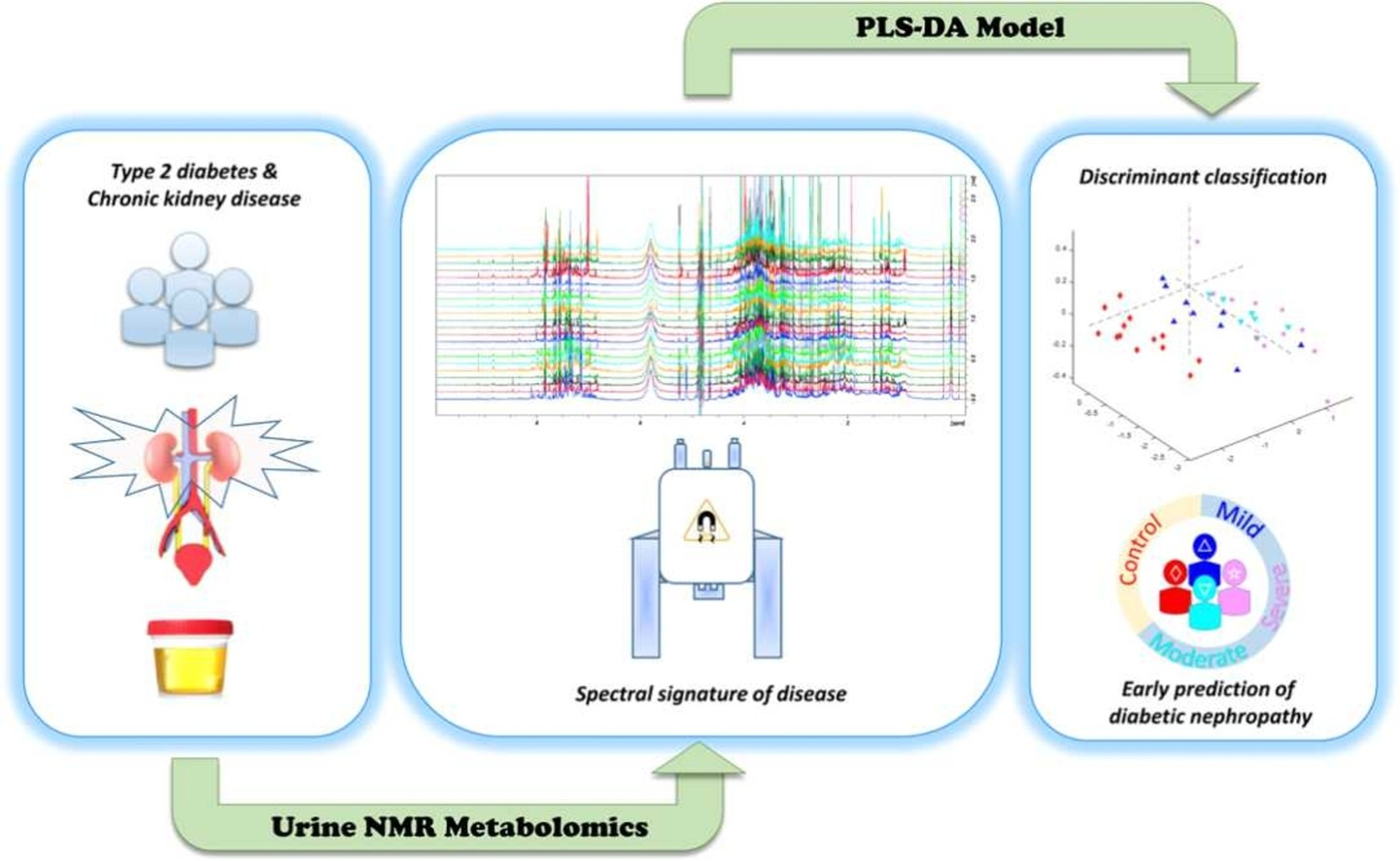
Type 2 diabetes mellitus (DM2) is a multimorbidity, long-term condition, and one of the worldwide leading causes of chronic kidney disease (CKD) –a silent disease, usually detected when non-reversible renal damage have already occurred. New strategies and more effective laboratory methods are needed for more opportune diagnosis of DM2-CKD. This study comprises clinical parameters and nuclear magnetic resonance (NMR)-based urine metabolomics data from 60 individuals (20–65 years old, 67.7% females), sorted in 5 experimental groups (healthy subjects; diabetic patients without any clinical sign of CKD; and patients with mild, moderate, and severe DM2-CKD), according to KDIGO. DM2-CKD produces a continuous variation of the urine metabolome, characterized by an increase/decrement of a group of metabolites that can be used to monitor CKD progression (trigonelline, hippurate, phenylalanine, glycolate, dimethylamine, alanine, 2-hydroxybutyrate, lactate, and citrate). NMR profiles were used to obtain a statistical model, based on partial least squares analysis (PLS-DA) to discriminate among groups. The PLS-DA model yielded good validation parameters (sensitivity, specificity, and area under the curve (AUC) of the receiver operating characteristic curve (ROC) plot: 0.692, 0.778 and 0.912, respectively) and, thus, it can differentiate between subjects with DM2-CKD in early stages, from subjects with a mild or severe condition. This metabolic signature exhibits a molecular variation associated to DM2-CKD, and data suggests it can be used to predict risk of DM2-CKD in patients without clinical signs of renal disease, offering a new alternative to current diagnosis methods.
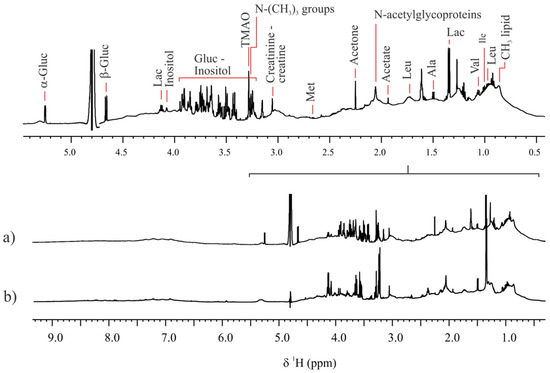
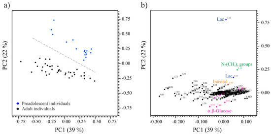
In goats, embryo oocyte competence is affected by follicle size regardless the age of the females. In previous studies we have found differences in blastocyst development between oocytes coming of small (<3 mm) and large follicles (>3 mm) in prepubertal (1–2 months-old) goats. Oocyte competence and Follicular Fluid (FF) composition changes throughout follicle growth. The aim of this study was to analyze Fatty Acids (FAs) composition and metabolomic profiles of FF recovered from small and large follicles of prepubertal goats and follicles of adult goats. FAs were analyzed by chromatography and metabolites by 1H-Nuclear Magnetic Resonance (1H-NMR) Spectrometry. The results showed important differences between adult and prepubertal follicles: (a) the presence of α,β-glucose in adult and no detection in prepubertal; (b) lactate, -N-(CH3)3 groups and inositol were higher in prepubertal (c) the percentage of Linolenic Acid, Total Saturated Fatty Acids and n-3 PUFAs were higher in adults; and (d) the percentage of Linoleic Acid, total MUFAs, PUFAs, n-6 PUFAs and n-6 PUFAs: n-3 PUFAs ratio were higher in prepubertal goats. Not significant differences were found in follicle size of prepubertal goats, despite the differences in oocyte competence for in vitro embryo production.
Representative 1H NMR spectra of follicular fluid samples of (a) adult and (b) prepubertal goats. Spectra were acquired at 298.0 K and at a magnetic field of 600 MHz, with suppression of the residual water signal.
(a) PCA scores plot (PC1-PC2) from 1H NMR spectral data of follicular fluid samples of prepubertal (n = 16; blue dots) and adult (n = 40; black dots) goats. (b) PCA heat map loadings plot (PC1-PC2) with some discriminant variables assigned.
Pulsed-Field Gradients (PFGs) play an important role in the development and understanding of modern NMR methods. With the ultimate goal of constructing robust pulse sequences that create high-quality NMR spectra with minimum set-up, PFGs are utilized to achieve an exclusive selection of a specific coherence transfer pathway as well as purging all kinds of undesired magnetization. PFG reduces the number of needed phase cycle steps to a bare minimum, allowing for accelerated NMR data acquisition in shorter spectrometer times. The potential and diversity of several PFG-based NMR elements are presented, as well as instances of their implementation in time-efficient NMR solutions. Practical aspects such as NMR data collecting needs and the attainment of pure in-phase absorption lineshapes are discussed for the most useful NMR experiments.
ISBN13: 9781839164002 Publisher: Royal Society of Chemistry Published: 17/05/2023
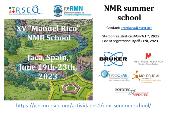
The Specialised Group of NMR of the Spanish Royal Society of Chemistry (GERMN, RSEQ) organizes the XV Manuel Rico NMR Summer School in Jaca from 19th-23th June 2023.
This well-established bi-annual summer course is aimed at PhD students, postdocs, technical staff of NMR facilities and, in general, to researchers from academy and industry interested in deepening their understanding of NMR. The course covers theoretical aspects, state-of-the-art methods and applications in fields as diverse as Molecular Chemistry, Materials, Biology, Medicine, and Pharmaceutical Industry, including solution-state, and solid-state NMR techniques, as well as MRI techniques.
Silvia Lope, SeRMN staff, will be teaching a class in “Magnetic Resonance Imaging”.
A fruitful collaboration between the Institute of Chemical Research of Catalonia (ICIQ) along with other international institutions involving the SeRMN-UAB has allowed the demonstration that a molecular magnetic memory near room temperature is a reality. A new way of storing information has come to light and possesses a potential technological impact. Molecular bits are already here.
Reference: Moneo-Corcuera, A., Nieto-Castro, D., Cirera, J., Gómez, V., Sanjosé-Orduna, J., Casadevall, C., Molnár, G., Bousseksou, A., Parella, T., Martínez-Agudo, J.M., Lloret-Fillol, J., Pérez-Temprano, M.H., Ruiz, E. & Galán-Mascarós, J.R. Molecular memory near room temperature in an iron polyanionic complex. Chem, 9, 373-393 (2023). https://doi.org/10.1016/j.chempr.2022.09.025
The Specialised Group of NMR of the Spanish Royal Society of Chemistry (GERMN, RSEQ) organizes the XIV Manuel Rico NMR Summer School in Jaca from 19th-24th June 2022.
This well-established bi-annual summer course is aimed at PhD students, postdocs, technical staff of NMR facilities and, in general, to researchers from academy and industry interested in deepening their understanding of NMR. The course covers theoretical aspects, state-of-the-art methods and applications in fields as diverse as Molecular Chemistry, Materials, Biology, Medicine, and Pharmaceutical Industry, including solution-state, and solid-state NMR techniques, as well as MRI techniques.
Silvia Lope, SeRMN staff, will be teaching a class in “Magnetic Resonance Imaging”.
End of pre-registration April 12th, 2022
Start of Registration: April 30th, 2022
End of Registration: May 17th, 2022
For detailed information please visit their webpage https://rmnjaca22.iqfr.csic.es/
Workshop limited to 4 participants (first come, first served)
Contact person:
Silvia Lope-Piedrafita, PhD ()
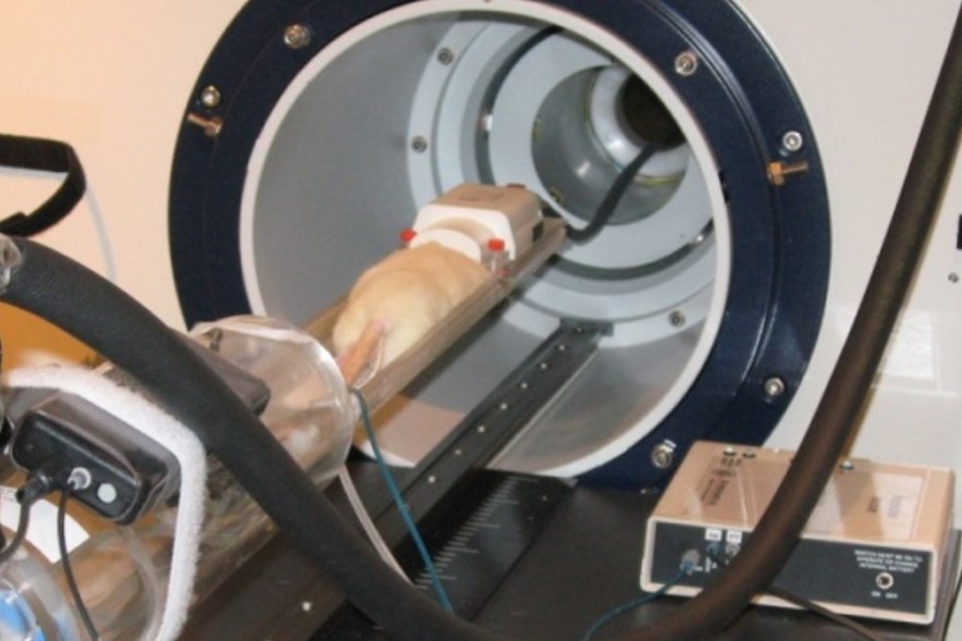
This course combines a comprehensive series of lectures on the technology of Magnetic resonance spectroscopy and imaging (MRS/MRI) with hands-on laboratory sessions to provide practical demonstrations of key concepts and procedures for preclinical studies.
Whether you are considering MRI as a research tool in your lab or just would like to learn more about MRI, this workshop addresses practical aspects of experimental MRI with laboratory animals and provide valuable hands-on experience on a 7 Tesla Bruker BioSpec spectrometer.